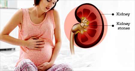
Kidney Stones and Pregnancy
When calcium combines with other substances in your urine, calcifications form that accumulate and block the passage of urine through the capillaries that carry urine from your kidneys to your bladder. These solid masses are known as kidney stones.
Kidney stones develop in pregnant women at a rate comparable to that of non-pregnant women of reproductive age, around one in per 1000 to 3000 pregnancies. By the age of 70, kidney stones harm 1 in 12 persons, 19% of males and 9% of women. 80 to 90 percent of stones occur in the second and third trimester, suggesting that multiparous women are more likely to have stones.
Symptoms of Kidney Stones in Pregnancy
- Blood in Your Urine a pregnant mother’s anxiety of miscarriage or other health issues may increase as hematuria develops, even if bloody urine from kidney stones is not a severe health risk in and of itself.
- Nausea or Vomiting might result from the pain and discomfort caused by kidney stones.
- Urinary Urgency and Frequency this is frequently an excruciating discomfort that makes urinating difficult.
- Lower Abdominal Pain/Sharp Pain in your Back this pain may last for a short or long time.
- Foul Smelling Urine urine might have alterations in its color and smell due to kidney stones.
Diagnosis of Kidney Stones in Pregnancy
- Assessment in the Laboratory: A midstream urine samples should be analyzed with a dipstick to check for underlying infections. An alkaline pH of greater than 7 may indicate urea-splitting organism infection, whereas a pH of less than 5 may indicate uric acid stone development. A urine culture and sensitivity test should be performed to confirm infection and identify the bacteria causing it if the dipstick results show nitrites. To check for anemia, renal function, and any electrolyte abnormalities, including calcium deficiency, blood should be sent. Examinations for hyperparathyroidism have to be prompted by elevated blood calcium levels.
- Radiological Diagnosis: which includes magnetic resonance imaging and magnetic resonance urography, carries dangers related to radiation that vary depending on the age of the pregnancy. In some circumstances, getting imaging using lower dosage methods to treat the stone is warranted. Since X-rays pose a greater risk to the fetus, they are strictly forbidden.
- Ultrasonography (US): Pregnant women who have been exposed to radiation have ultrasonography as their first line of examination because of the known hazards to the developing fetus. Sensitivities for stone detection, however, range widely, from 29 to 69%. Differentiating between pathologic and normal hydronephrosis can be challenging, despite the fact that US offers more details on hydronephrosis and the hydroureter. It’s possible that 20% of patients who have total blockage go unnoticed because they are attributed to “physiological hydronephrosis” during pregnancy. It is significant to note that ureteral dilatation in physiological hydronephrosis typically does not extend past the iliac artery and below the pelvic brim. The diagnosis of ureteral calculi was successfully ruled out in research comparing the use of US and renography in pregnant women with hydronephrosis when the renal pelvic diameter in asymptomatic patients was less than 17 mm.
- Intravenous urography or CT scanning: In cases where ultrasonography is unable to identify stones and symptoms such pain, vomiting, and fever persist, or if renal function is declining, other diagnostic techniques can be taken into account. On which is better, there isn’t a certain consensus. One alternative that has been suggested is limited IVU. Sensitivities for the various restricted IVU methods that have been proposed range from 60 to 94%.
Some have argued for the use of low-dose CT in cases where US has proved inconclusive, despite the fact that unenhanced CT is normally not advised during pregnancy due to increased ionizing radiation exposure. In a retrospective research, renal US and low-dose CT scan were performed on 20 pregnant women suspected of having urolithiasis. In comparison to renal US, the study indicated that CT was more sensitive in identifying urinary calculi. Compared to the conventional mean of 25 mGy for CT pelvis, the radiation dosage utilized ranged from 2 to 13.7 mGy. They came to the conclusion that, in some cases, sparingly applying low-dose CT might enhance patient care.
- MRI urography: MRI does not employ ionizing radiation; instead, it uses electromagnetic radio waves. There have been no known negative effects on the fetus, but because there isn’t enough information on its safety during fetal organogenesis, it should be avoided throughout the first trimester. MRU is the use of MRI to see the urine system. Unenhanced, strongly T2 weighted pulsed signals have been utilized in studies to produce pictures where static fluid has a larger signal intensity compared to the background (static MRU). To get information on the kidneys excretory function, MRU can also be done with a contrast agent, such as gadolinium (excretory MRU).
Treatment of Kidney Stones in Pregnancy
Depending on the size of the stone, there are many treatment options for kidney stones. Furthermore, pregnancy may limit the kinds of treatments your doctor suggests for your particular situation.
- Using Sound Waves to Shatter Stones
The purpose of this treatment is to target stones that have significant vibrations that can lessen their size and allow them to pass. Even though it is non-invasive, some pregnant women may want to avoid the uncomfortable and perhaps sedation-requiring process.
- Procedure Scope
Using a scope to break up the stone by passing it up your urethra and into your ureters might be a possibility for smaller stones. But as you recuperate, you could need a stent, and the surgery itself can call for anesthesia.
- Medication for Pain
In cases when kidney stones have obstructed the ureters, doctors may give specific muscle relaxants to ease the obstruction. Painkillers available over-the-counter or with a prescription can be used to treat kidney stone pain.
Medication may be recommended to stop the creation of new stones, depending on the kind of stone. These might include drugs to regulate uric acid, oxalate, or calcium levels.
- Lithotripsy with Laser
In order to make the kidney stone simpler to pass or remove the fragments by other treatments, a laser is utilized to split it up into tiny pieces.
- Surgery
Surgery to remove the parathyroid gland may be considered in situations when the glands overactivity is causing stones to develop.
Ways to Treat Kidney Pain during Pregnancy
- Drinking plenty of water (It’s usually advised to drink lots of water to help dissolve the stone and stop it from forming again. Staying well hydrated is essential for avoiding kidney stones). Drinking enough water each day—roughly two to three quarts, or ten 10-ounce glasses—can prevent more stones. Keep in mind that drinking more water can help you replenish the fluids you lose via perspiration during physical activity or in hot conditions. Drinking largely calorie-free or low-calorie beverages is ideal. Reducing alcohol or coffee-based beverages may be necessary for this.
- Consult Your Doctor About Reducing Your Intake of Calcium Supplements (Although some pregnant women are advised to take calcium supplements, there is a chance that this is raising the risk of kidney stones).
- Warm Baths (Taking a warm bath might assist in relieving pain and relaxing muscles. In particular when pregnant, make sure the water is not too hot).
Conclusion
Treating kidney stones as soon as possible is essential to reducing the chance of further health issues. To receive a precise diagnosis and create a treatment plan that takes your condition into consideration while putting the health of your unborn child first, speak with your doctor right away.